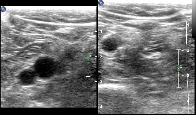

The configuration of the valves at the SFJ is important to consider as there is typically a terminal valve located at the junction and a preterminal valve that is positioned in the GSV, 3–5 cm distal to the terminal valve. Finally, there are usually intersaphenous veins in the upper calf, running between the GSV and SSV.įigure 13.10 An image of a venous valve site in the popliteal vein just below the gastrocnemius vein. Generally, the Giacomini vein should appear to run into an intrafascial compartment in the lower thigh before joining the SSV, whereas superficial tributaries of the GSV tend to lie above the superficial fascia. There is sometimes confusion as to whether a posterior thigh vein is the Giacomini vein or merely a superficial posteromedial tributary of the GSV. The TE is often referred to as the Giacomini vein, although strictly this term should only be applied when the vein connects to the GSV. There are a number of terminations for the TE, as shown in Figure 13.8. Note that it is possible to confuse the posteromedial tributary of the great saphenous vein with the Giacomini vein see text.


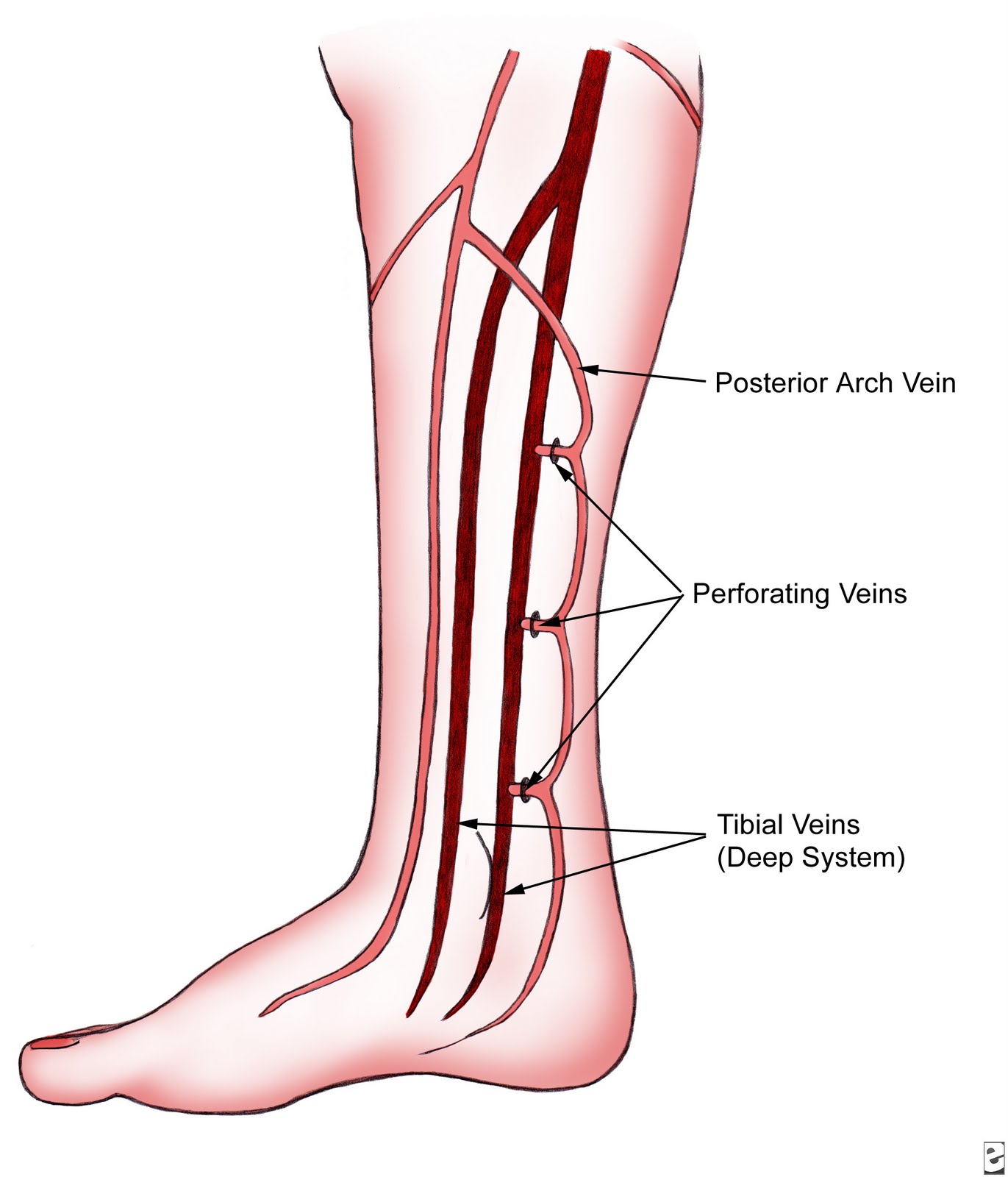
It may also drain to the great saphenous vein (GSV) and in this situation the vein is termed the Giacomini vein (orange line). It may drain to the femoral vein, profunda femoris vein or branches of the internal iliac vein (green lines). The thigh extension vein, when present, runs above the level of the saphenopopliteal junction and has a variable outflow. It sometimes has a high junction with the popliteal vein (blue dashed line). It can share a common junction with the gastrocnemius vein (diagram C). It can share a common trunk (CT) with the gastrocnemius vein (GV) (diagram B). The SSV normally drains to the popliteal vein (P) in the popliteal fossa at the saphenopopliteal junction (diagram A). Potential positions are shown by numbers and lettered diagrams. The left common iliac vein runs deep to the right common iliac artery to drain into the vena cava, which lies to the right of the aorta.įigure 13.8 The level of the small saphenous vein (SSV) insertion can be highly variable. The external iliac vein runs deep and is joined by the internal iliac vein, which drains blood from the pelvis, forming the common iliac vein. The common femoral vein lies medial to the artery, becoming the external iliac vein above the inguinal ligament ( Fig. This confluence is distal to the level of the saphenofemoral junction (SFJ) and common femoral artery bifurcation (see Fig. The femoral vein runs toward the groin, where it is joined by the deep femoral vein to form the common femoral vein. The above-knee popliteal vein runs through the adductor canal and becomes the femoral vein in the lower medial aspect of the thigh. The anterior tibial vein is paired and associated with the anterior tibial artery and drains to the popliteal vein. The gastrocnemius veins normally drain into the popliteal vein through single or multiple trunks below the level of the saphenopopliteal junction. The gastrocnemius veins drain the medial and lateral gastrocnemius muscles. They are an important part of the calf muscle pump mechanism (see Ch. The soleal veins are deep venous sinuses and veins of the soleus muscle that drain into the popliteal vein. The paired veins join into common trunks in the upper calf before forming the below-knee popliteal vein. The posterior tibial and peroneal veins are usually paired and are associated with their respective arteries. This vein is part of the deep venous system. The term superficial is misleading and may be misinterpreted. The Union Internationale de Phlébologie recommends the use of the term femoral vein instead of superficial femoral vein to describe the vein running between the common femoral vein and popliteal vein.


 0 kommentar(er)
